What Is Pleural Mesothelioma?
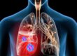
Pleural mesothelioma is a type of cancer caused by asbestos fibers becoming embedded in the lining of the lungs. Over time, the fibers may cause inflammation and scarring. As the scarring worsens, it may develop into mesothelioma tumors.
- Pleural malignant mesothelioma is the most common form of mesothelioma cancer.
- Each year, about 2,500 people are diagnosed with the disease.
- Symptoms of the cancer commonly include shortness of breath, chest pain, dry cough and fatigue.
- Diagnosis typically consists of multiple tests, including scans and biopsies.
- Pleural mesothelioma is often treated with chemotherapy, surgery, radiation therapy and immunotherapy.
- A patient’s prognosis will vary depending on their individual case, with an average life expectancy of about 18 months.

Prognosis
A mesothelioma prognosis is what doctors describe as an overall outlook for a specific patient. Mesothelioma is aggressive cancer with an average life expectancy of 14 to 22 months, but it is often terminal with most patients living only 1 year after diagnosis. A patient’s prognosis helps determine the treatment options they can pursue.
The most important factors affecting the prognosis of pleural malignant mesothelioma patients are:
- Cell type (histopathology)
- Stage of the cancer
- Patient’s gender and age
Three Types of Mesothelioma Cells
- The three mesothelioma cell varieties are epithelial, sarcomatoid, and biphasic. Biphasic is a mix of the first two cell types.
- Mesothelioma doctors can tell the difference between cells based on how they look under a microscope.
- Different mesothelioma tumors respond differently to a treatment. Epithelial or epithelioid cells respond the best, and sarcomatoid cells are more resistant to treatment.
- Cancer doctors take these differences into account when planning your mesothelioma treatment. The incidence of these cell types varies by cancer location.


Prevalence of Pleural Mesothelioma Tumors by Cell Type
| CELL TYPE | PERCENT |
| Epithelioid | 50% |
| Biphasic | 30% |
| Sarcomatoid | 20% |
What Are the Symptoms of Pleural Mesothelioma?
Pleural mesothelioma, which affects the tissue that surrounds the lungs, causes signs and symptoms that may include:
- Chest pain.
- Painful coughing.
- Shortness of breath.
- Unusual lumps of tissue under the skin on your chest.
- Unexplained weight loss.
Pleural malignant mesothelioma patients may be diagnosed with a co-occurring asbestos-related condition, which can impact symptom onset. These include:
- Pleural plaques – Chalky substance that forms on the pleura due to a buildup of minerals, known as calcification
- Diffuse pleural thickening (DPT) – Gray, fibrous tissue that fills in pleural spaces
- Asbestosis – Scarring of the lungs (fibrosis)
How Is Pleural Mesothelioma Diagnosed?
- Symptoms: Symptoms of mesothelioma cause the patient to visit their primary care physician or a hospital for testing.
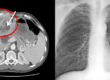
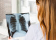
- Mesothelioma Testing: The most common first tests are radiology exams such as chest X-rays and CT scans of the chest or abdomen. These initial tests reveal abnormal results, which lead to further testing and referral to a surgeon or oncologist.
- Confirm Diagnosis: The surgeon or oncologist uses a combination of imaging scans, blood tests, and biopsies to confirm a mesothelioma diagnosis. A blood test or radiology scans alone cannot confirm a diagnosis of mesothelioma.
İmaging Tests Commonly Used in Mesothelioma Diagnosis
- X-Rays: Produce basic images of areas with various densities, such as tumors or fluid, within the body.
- CT Scans: Also known as CAT scans. These use computer software to integrate hundreds of fine X-ray images to create detailed images of internal structures. These images are much more precise than regular X-rays.
- MRIs: Using electromagnetic technology, MRIs generate precise images, which are especially useful when looking at bone, nerve and brain tissue.
- PET Scans: These are CT scans where the patient receives an intravenous dose of radioactive glucose, which makes inflamed cells light up on scans. Cell inflammation may be caused by an infection or rapid growth, which could be a sign of cancer.
There are three mesothelioma biopsy types that your doctor may consider:
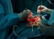
- Surgical biopsies are the most invasive and involve removing a large sample of the mesothelioma tumor or the whole tumor.
- Endoscopic biopsies use a thin tube with a tiny camera on the end to examine the affected tissue and take a sample for testing.
- Needle biopsies involve using a very thin needle to remove a sample of the tumor or fluid from the chest cavity, abdominal cavity, or heart sac.
Endoscopic Biopsies
Endoscopes are slender, tube-shaped instruments with a small video camera and light on the tip to help guide them. Your doctor may use an endoscopic biopsy to examine spots inside the body and take tissue samples.
- Thoracoscopies
Thoracoscopies are used to examine and take samples of the pleural tissue lining the lungs and chest cavity. This procedure can also be used to take samples of lymph nodes in the area to check on whether the cancer is spreading.
- Laparoscopies
Laparoscopies allow your doctor to examine the abdominal cavity and take tissue samples of any tumors.
- Mediastinoscopies
Mediastinoscopies are used to take samples from the space between the lungs, known as the mediastinum. This is an area that contains lymph nodes, which your doctor may want to biopsy to determine whether the cancer has spread.
- Bronchoscopies
Bronchoscopies may also be used in some cases to obtain samples of lymph nodes near the lungs.
- Endoscopic procedures require general anesthesia and must be done in the operating room, but they are not as invasive as surgical biopsies.
Needle Biopsies
There are several types of needle biopsies, often referred to as fine needle aspirations, that may be used to help diagnose mesothelioma. For these tests, the doctor will numb a patch of your skin and then insert a thin needle. Some needle biopsies remove fluid that has built up in the body due to the cancer, and others take a sample of the tumor itself or nearby lymph nodes.
- Thoracentesis
Thoracentesis is used to take a sample of fluid that has built up in the chest (a condition known as pleural effusion). This procedure can also be used to help ease a patient’s pain from the fluid build-up, which is a common symptom of pleural mesothelioma.
- Paracentesis
Paracentesis is used to take a sample of fluid that has built up in the abdomen (a condition known as ascites). It can also help relieve a patient’s discomfort in the abdominal area, a common symptom of peritoneal mesothelioma.
- Pericardiocentesis
Pericardiocentesis is used to take a sample of fluid from the pericardial sac surrounding the heart. This type of fluid build-up is common in pericardial mesothelioma patients.
All of these outpatient procedures can be done at your doctor’s office or in a hospital setting. Your doctor may use imaging tests such as a CT scan or ultrasound to help direct the needle. Although these are the least invasive types of mesothelioma biopsies, the results may not be conclusive enough to make a mesothelioma diagnosis.
Pleural Mesothelioma Treatment
Pleural mesothelioma is typically treated with a multimodal approach, combining standard treatments like surgery, chemotherapy, and radiation. A pleural mesothelioma treatment plan will largely depend on the cell type and stage of the disease. Generally, treatment plans are not intended to cure the disease.
Mesothelioma surgery is common for pleural mesothelioma patients. The surgery may be aggressive with the goal of extending life expectancy, or less aggressive with the goal of palliating symptoms (relieving discomfort).
Surgery may be an option for early-stage malignant pleural mesothelioma patients. Pleurectomy/decortication (P/D) is one common surgical option that involves the removal of the lining of the lung and chest wall, as well as other impacted tissues and organs.
Another option is extrapleural pneumonectomy (EPP). The more aggressive procedure includes removing the affected lung, part of the diaphragm, and the linings of the heart and lungs. Recent clinical trials have found these surgeries can extend life expectancy to three years or longer, especially when applied multimodally with chemotherapy and/or radiation.


References
- https://www.mesothelioma.com/mesothelioma/types/pleural/
- https://www.asbestos.com/mesothelioma/diagnosis/
- https://www.mesotheliomahelp.org/mesothelioma/diagnosis/biopsy-types/
- https://en.wikipedia.org/wiki/Mediastinal_tumors
- https://www.annalscts.com/article/view/1054/1583