Introduction
Thoracic trauma accounts for up to 35% of trauma-related deaths in the United States and encompasses a broad range of injuries that can cause significant morbidity and mortality (1).
Prompt evaluation during the primary trauma survey is key to identifying those injuries which are immediately life-threatening and require rapid intervention. Once these conditions are ruled out, less urgent thoracic injuries are often readily diagnosed during the secondary trauma survey and successfully managed by applying the fundamental principles of advanced trauma life support (ATLS).
Etiology
Thoracic trauma is broadly categorized by mechanism into blunt or penetrating trauma. The most common cause of blunt chest trauma is motor vehicle collisions (MVC) which account for up to 80% of injuries.
Other causes include falls, vehicles striking pedestrians, acts of violence, and blast injuries. The majority of penetrating trauma is due to gunshots and stabbings, which together account for 20% of all major trauma in the United States (2).
Pathophysiology
Most morbidity and mortality due to chest trauma occurs because injuries interfere with respiration, circulation, or both.
Respiration can be compromised by
- Direct damage to the lungs or airways
- Altered mechanics of breathing
Injuries that directly damage the lung or airways include pulmonary contusion and tracheobronchial disruption. Injuries that alter the mechanics of breathing include hemothorax, pneumothorax, and flail chest. Injury to the lung, tracheobronchial tree, or rarely esophagus may allow air to enter the soft tissues of the chest and/or neck (subcutaneous emphysema) or mediastinum (pneumomediastinum). This air itself rarely has significant physiologic consequence; the underlying injury is the problem. Tension pneumothorax impairs respiration as well as circulation.
Circulation can be impaired by
- Bleeding
- Decreased venous return
- Direct cardiac injury
Bleeding, as occurs in hemothorax, can be massive, causing shock (respiration is also impaired if hemothorax is large). Decreased venous return impairs cardiac filling, causing hypotension. Decreased venous return can occur due to increased intrathoracic pressure in tension pneumothorax or to increased intrapericardial pressure in cardiac tamponade. Heart failure and/or conduction abnormalities can result from a blunt cardiac injury that damages the myocardium or the heart valves.
Common Complications Following Thoracic Trauma
Although there are a wide range of complications following thoracic trauma,
- Respiratory failure,
- Pneumonia, and
- Pleural sepsis is the most common potentially preventable problem.
Respiratory failure and pneumonia are directly related to the severity of the injury and the age and condition of the patient. A program aimed at aggressive pain control, mobilization, and pulmonary care can reduce the risk of respiratory failure, pneumonia, and death in these patients.
Pleural sepsis develops in the face of a retained hemothorax, which becomes contaminated with bacteria.
What are the symptoms of chest injuries?
Signs of chest injury can vary, depending on the type of injury. The most common signs and symptoms are:
- Pain in the chest that gets worse when laughing, coughing, or sneezing
- Tenderness
- Bruising
- Swelling
Symptoms of a fractured rib are:
- Extreme pain when breathing in
- Tenderness to the chest or back over the ribs
- A ‘crunchy’ feeling under the skin
- Severe shortness of breath
How are chest injuries diagnosed?
A chest injury is diagnosed with a physical examination and sometimes investigations such as a chest x-ray. A blood test may also be done. A CT scan may also be needed to check for injury to the heart.
For a rib fracture, sometimes doctors can feel the broken ribs when they gently press the affected area. Sometimes rib fractures don’t show on a chest x-ray. If you appear well and the doctor doesn’t suspect complications, you may not need an x-ray. Your doctor may order a chest x-ray to look for any serious problems related to the fractured rib, such as a bruised or collapsed lung.
Clinical evaluation
Five conditions are immediately life threatening and rapidly correctable:
- Massive hemothorax (Blood in the space surrounding the lung)
- Tension pneumothorax (Trapped air in the space surrounding the lung)
- Open pneumothorax (Air in the space surrounding the lung)
- Flail chest (A life-threatening medical condition that occurs when a segment of the rib cage breaks due to trauma and becomes detached from the rest of the chest wall. Two of the symptoms of flail chest are chest pain and shortness of breath).
- Pericardial tamponade (A serious medical condition in which blood or fluids fill the space between the sac that encases the heart and the heart muscle. This places extreme pressure on your heart)
- Surgical emphysema (Air trapped under the skin can cause a swollen area on the chest Wall)
- Abdominal injuries (Liver or spleen damage can cause pain in your stomach or back)
Diagnosis and treatment begin during the primary survey and are based first on clinical findings. Depth and symmetry of chest wall excursion are assessed, the lungs are auscultated, and the entire chest wall and neck are inspected and palpated. Patients in respiratory distress should be monitored with serial assessments of clinical status and of oxygenation plus ventilation (eg, with pulse oximetry, arterial blood gas tests, capnometry if intubated).
Penetrating chest wounds should not be probed. However, their location helps predict risk of injury. High-risk wounds are those medial to the nipples or scapulae and those that traverse the chest from side to side (ie, entering one hemithorax and exiting the other). Such wounds may injure the hilar or great vessels, heart, tracheobronchial tree, or rarely the esophagus.
Patients with symptoms of partial or complete airway obstruction following blunt trauma should be immediately intubated to control the airway.
In patients with difficulty breathing, severe injuries to consider during the primary survey include the following:
- Tension pneumothorax
- Open pneumothorax
- Massive hemothorax
- Flail chest
Treatment / Management
How are chest injuries treated?
Treatment of the chest injury will depend on the cause of the injury and how serious it is. The medical team will support breathing and circulation if necessary. You may be given oxygen and intravenous fluids or blood transfusions. If you have a severe chest injury, you will be admitted to the hospital.
A fractured rib will heal on its own, but it takes time. If you have a fractured rib, you may be asked to breathe deeply regularly to keep the air sacs in the lung open and prevent pneumonia, a type of chest infection.
If you are in pain, take pain killers. Pain relief is important so you will feel more comfortable to cough and take deep breaths.
If your injury is minor, try to keep moving around and doing what you normally do. But avoid lifting, bending and any strenuous exercise until your pain and other symptoms have gone.
Initial Rapid Chest Injury Treatment Steps
- Begin CPR, if Necessary.
- Cover an Open Wound.
- Stop Bleeding, if Necessary.
- Position Person to Make Breathing Easier.
- Monitor Breathing.
- Follow Up.
After a primary survey immediately life-threatening injuries should be excluded or treated such as:
- Airway obstruction;
- Tension pneumothorax;
- Open pneumothorax;
- Massive haemothorax;
- Flail chest;
- Cardiac tamponade.
Secondary survey will provide information on potentially life-threatening injuries:
- Pulmonary contusion;
- Myocardial contusion;
- Aortic disruption;
- Traumatic diaphragmatic rupture;
- Tracheobronchial disruption;
- Oesophageal disruption.
Life-threatening injuries diagnosed during the initial trauma evaluation require prompt intervention. Still, the most common injuries due to thoracic trauma are pneumothorax and hemothorax which are definitively managed in 80% of cases with tube thoracostomy. The size of the chest tube used is a clinical decision based on the pathology seen on a chest x-ray. If both pneumothorax and hemothorax are present a size 28-Fr or 32-Fr chest tube is usually considered as this will facilitate the evacuation of both air and blood while minimizing the chance of the tube obstructing due to clot. If no effusion is present, small-bore catheters are appropriate although many trauma clinicians will still opt for formal chest tubes instead. Occult pneumothorax is a pneumothorax which is seen on CT but not on a chest x-ray. They are incidentally found in 2% to 10% of trauma patients who undergo chest CT. Patients can be observed if the pneumothorax is less than 8 mm (3).
However, occult pneumothoraces are associated with a 5% to 10% risk of expansion and should, therefore, be monitored closely. Patients whose pneumothoraces expand or those who become symptomatic warrant tube thoracostomy.
When should the thoracic surgeon definitely be involved?
According to the ATLS guideline this is recommended as follows (4):
- Blood loss over the chest TD >1,500 mL initially or >200 mL/hour over 2–4 hours;
- Haemoptysis;
- Massive subcutaneous emphysema;
- Important air-leakage over the chest tub;
- Uncertain images on the chest X-ray or CT thorax;
- Penetrating chest trauma.
Indications for an immediate thoracic surgical intervention are (4):
- Blood loss ≥1,500 mL initially/>200 mL/hour over 2–4 hours;
- Endobronchial blood loss; massive contusion with significant impairment of mechanical ventilation;
- Tracheobronchial tree injury (air-leakage/hemothorax);
- Injury of the heart or large vessels (blood loss/pericardial tamponade).
Surgical trauma life support
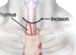
Which incision should be chosen for emergency thoracic surgical intervention? Anterolateral thoracotomy in the 4–6th intercostal space is usually recommended, although in 20% of the patients it is insufficient to visualize all lesions and must therefore modified (5).
Clamshell (transverse sternotomy and bilateral anterolateral thoracotomy) or Hemi-clamshell (longitudinal sternotomy and anterolateral thoracotomy) will permit better exposition of thoracic organs. The necessity for emergency room thoracotomy is extremely rare, anterolateral thoracotomy will permit a potentially lifesaving measure (clamping of a great vessel) in an extreme situation before proceeding to the operating theatre (6).
A thoracoscopic evaluation of the pleural cavity can demonstrate easily misdiagnosed lesions and treat a possible persisting haemothorax. It is obvious that VATS (Video-assisted Thoracic Surgery) can play a role in the treatment of chest trauma management so long the inclusion criteria are respected. In a haemodynamic unstable patient with severe chest wall or cardiac vessels injuries and massive transfusion there is no place for thoracoscopic efforts that only delay the unavoidable open approach and perhaps minimize the chances for a positive outcome. But in the stable haemodynamic state with small perforating wounds, VATS can be a valuable weapon of thoracic surgeons for fast recovery, minimized pain and perfect visualization of the entire pleural place. Indications for such an approach are as followed:
Indications for VATS in severely injured patients:
- (Penetrating) injury with little blood loss in a stable patient;
- Persistent hemothorax;
- Empyema;
- Persistent air-leakage;
- Suspicion of diaphragmatic rupture.
Conclusions
Trauma care is complex. Blunt thoracic trauma is frequent but emergency surgical interventions are rare. Thoracic surgeons are usually not part of the trauma team in most trauma centres. The objective was to review specific details in thoracic trauma care, is there a place for minimal invasive surgery in thoracic trauma care and to demonstrate how the experience of the thoracic surgeon may be of advantage for severely injured patient.
References
- Surg Clin North Am 2007 Feb;87(1):95-118, vii.
- J Trauma 1990 Nov;30(11):1356-65.
- J Trauma 2011 May;70(5):1019-23; discussion 1023-5.
- J Trauma Acute Care Surg 2013;74:1363-6.
- J Trauma 2004;56:664-8.
- J Trauma 2004;57:576-81.